|
Liquid ionization chambers are developed to be used as monitors in radiation
therapy. The development focuses on two areas of application, an on-line
monitor for intensity modulated radiation therapy and the measurement of the
energy deposit of heavy ions in tissue. This requires a thin detector with
two-dimensional read-out and an absorbing chamber with three-dimensional
read-out, respectively.
The work is done in collaboration with the German Cancer Research Center
(DKFZ) in Heidelberg (Prof. Dr. G. Hartmann).
|
|
|
|
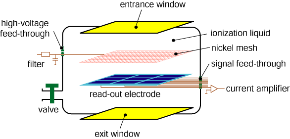
Both detectors are realized using similar techniques. Ionizing radiation is
measured with liquid ionization chambers, which are operated at room
temperature. The liquids isooctane, isononane (TMP), and tetramethylsilane
(TMS) are used in a high purity grade in order to realize high current signals.
Finely segmented read-out electrodes with typically several hundred individual
channels are utilized. They are read out in parallel at frequencies exceeding
10 Hz.
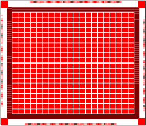
The figure depicts a read-out electrode with 400 channels.
|
|
|
|
Chamber with two-dimensional read-out
|
|
|
A chamber with two-dimensional read-out has been designed to test the liquids
and to gain experience with the principal components of an envisaged monitor
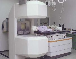
chamber. The dimensions and the frame have been chosen to be used gantry
mounted with a miniature multileaf collimator (ModuLeaf, MRC Systems GmbH,
Heidelberg, Germany) and a X-ray beam of a linear accelerator (PRIMUS, 6/15
MV, Siemens OCS, Concord, California, USA) at the DKFZ. The aperture of the
collimator is 70 x 84 mm2. The entrance and exit windows are of
special inert plastic material (VECTRA), which with its low mean atomic number
is sufficiently transparent for X-rays and is flexible enough to allow for
temperature and pressure variations.
The picture shows the prototype before being filled with ionization liquid.
|
|
|
|
The distance between the electrodes has been chosen to 5 mm in order to
maximize the signal and to minimize the electronic noise. The voltage (up to 5
kV) is applied to the cathode which consists of a thin nickel mesh. The pad
structure of the anode is realized as a silver palladium conductor on a ceramic
substrate. The pad rows are aligned with the collimator leafs and the pad size
of 3.3 x 4.0 mm2 and the spacing of 0.2 mm is adapted to the leaf
width of 1.75 mm. The positions of two adjacent leafs are controlled by one row
of pads. The 400 pads are surrounded by a guard electrode (3.5 mm wide
rectangular frame) in order to form at the edges an adequate homogeneous
electric field.
The figure illustrates an electrode configuration for a chamber with
two-dimensional read-out.
|
|
|
|
Electronically the anodes are on virtual ground and the field shaping
electrode is on ground potential. Each pad is connected to a circuit track on
the rear side of the ceramic substrate. The photo
shows the circuit track system which traces the signals to four
feed-through plugs. The tracks have a width of 125 micron and approach each
other 250 micron at most.
|

|
|
|
The chamber with two-dimensional read-out has been tested successfully using
beams from a 60Co source and an electron linear accelerator at the
DKFZ.
The current image of an asymmetrical cross formed with a multi leaf collimator
is shown. The insert represents the collimator aperture: the broad arms are 7
mm wide (corresponding to 4 leafs) and the thin arms have a width of 3.5 mm.
In conclusion, it can be stated that the chamber exhibits stability of
performance and shows uniformity in sensitivity and read-out.
|

|
|
|
Chamber with three-dimensional read-out
|
|
|
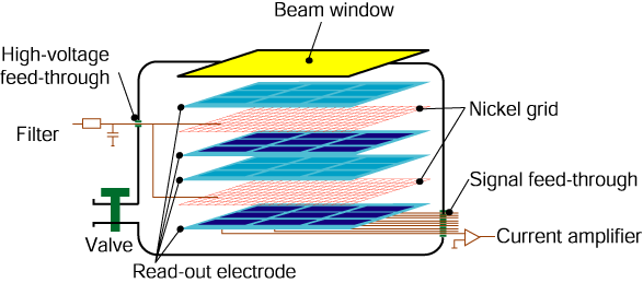
The ionization liquids used exhibit similar physical properties as human
tissue. Therefore, a liquid ionization chamber can be used to measure the
penetration of the radiation into the body in a phantom.
The measurement of the energy deposit as function of the depth in a medium
requires a monitor with three-dimensional read-out. To test this approach a
chamber with four planes of read-out electrodes has been built. Each layer
contains 10 x 10 pads with the dimensions 2.5 x 2.5 mm. Two pairs of anode
layers are arranged as sketched in the figure. In each layer pair the anode
pads are facing each other. The high voltage is applied to two nickel grids,
arranged in the center plane of each pair of anode layers. The distance between
the nickel grid and the read-out layers has been chosen to be 4 mm.
|
|
|
|
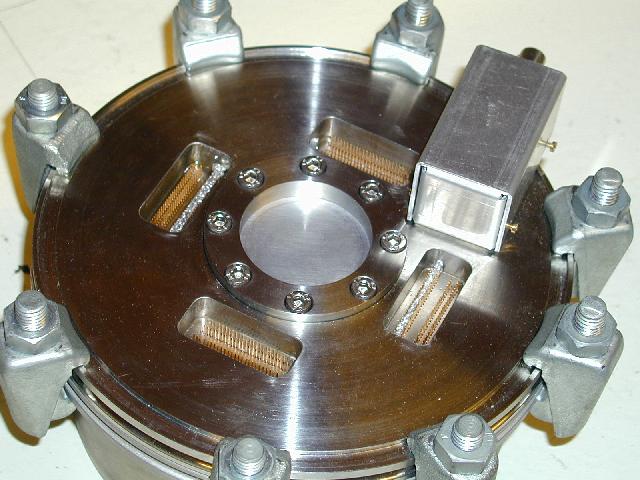
The materials used and the read-out electronics are identical to the set-up of
the chamber with two-dimensional read-out.
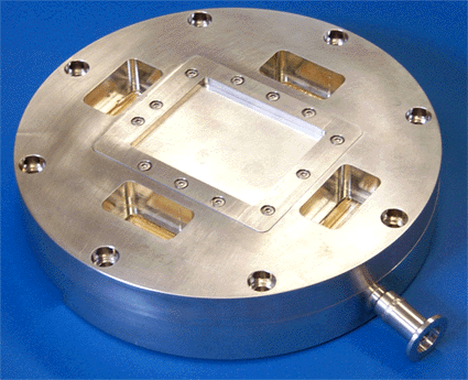
The photograph depicts the chamber with the four vacuum feed-troughs for the
signals of the 400 individual channels, arranged around the beam entrance
window in the center of the chamber lid.
in the
|
|
|
|
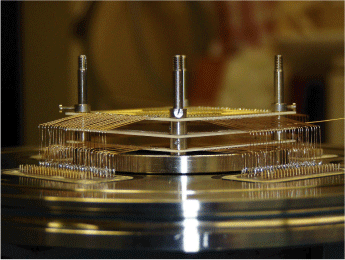
The photograph shows the set-up for the first two layers of the chamber
with three-dimensional read-out.
First tests of this chamber have been conducted at a 60Co-source at
the DKFZ in Heidelberg. It could be demonstrated that a homogeneous read out
can be achieved with this device. To verify the capability to measure the Bragg
peak it is planned to test this chamber at a 12C-beam at the GSI in
Darmstadt.
|
|
|